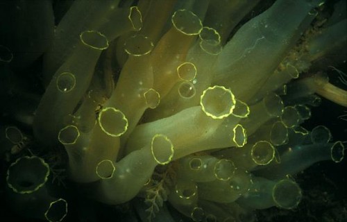
Dr. Vincent Pieribone, Associate Professor of Neurobiology at the Yale School of Medicine, is currently submarine diving in the Solomon Islands in search of shining proteins. If he finds them, it will represent a major advance in the field of neurology.
Understanding how the brain works is undeniably one of the greatest challenges of our time, and one that stretches back hundreds of years. Although scattered hypotheses have long strained for a coherent theory of the brain, efforts have been limited by our ignorance of the brain’s labyrinthine circuitry. “Traditionally, we would rely on invasive electrodes to sample very limited areas of the brain, and only a few neurons at a time,” Pieribone explained. This was characterized in a 2012 paper in the journal Neuron as viewing “an HDTV program by looking just at one or a few pixels on a screen.” Imaging technologies such as functional magnetic resonance imaging (fMRI) have improved our historical myopia of the brain’s inner circuits, but they are categorized as indirect methods which monitor areas of high metabolism in the brain, as opposed to actual neurons firing.

A collaboration between Dr. Vincent Pieribone and Dr. Michael Nitabach of the Yale School of Medicine has produced a chimeric, engineered molecule which has the power to illuminate the circuitry of the brain. The molecule, dubbed ArcLight, has two functional components. The first is a voltage sensor domain, a transmembrane protein module containing positively charged amino acids. Voltage changes across the membrane, which occurs when neurons fire, cause a change in the electric field in which these positive charges sit, and that imposes a force on the positive charges and changes the conformation of the domain. The second component, a mutated form of green fluorescent protein (GFP), is fused to the voltage sensor domain. When the sensor domain changes conformation, it also changes the shape of the GFP, which affects the level of fluorescence. Thus, ArcLight can be used to effectively map millions of neurons firing in real time by the intensity of fluorescence each gives off. Results demonstrating the efficacy of this method in fruit flies was published in the journal Cell in August.

The implications of this discovery are striking. “Behaviors and biological processes can be associated with patterns of light,” explained Pieribone. “For example, when people touch something, there is a pattern associated with that action. We can ask: what does the brain do? Or, to add a twist, what happens if you lose your arms?” In their study, Nitabach and Pieribone used ArcLight to demonstrate that neurons involved in the circadian rhythm of a fruit fly were more active in the morning than the evening. “This is something you could only see this way,” said Nitabach.

However, even this new method of neural imaging is not without limitations. Current efforts to improve the protein probe include increasing the intensity of the fluorescent signal in response to smaller voltages and increasing the speed of response (the current lag time is about 20 milliseconds). Moreover, the GFP is limited to absorbing blue light and emitting green light. If alternate versions could be discovered that can absorb green light and thus emit red light, ArcLight could be used in conjunction with other fluorescent sensors. One example would be calcium detectors which emit green light, allowing researchers to produce a system to investigate other variables of brain activity. Finding these alternate versions is one of the reasons why Pieribone is diving in the deep Pacific. If the new proteins are found, neural systems in which specific processes can be excited by light using a technique called optogenetics can be used concomitantly with ArcLight in order to selectively and comprehensively decode the electrical circuits that lie at the heart of neuroscience.


Last April, the Obama administration unfurled a long term scientific effort to build a comprehensive map of the human brain and its activity in the Brain Research through Advancing Innovative Neurotechnologies (BRAIN) Initiative. The initiative is a critical investment in executive eyes, projected at 300 million dollars per year for the next ten years. Here at Yale, in the labs of Nitabach and Pieribone, it looks like it is already paying off.
