There are more than twenty thousand different proteins in your body, and each one plays some role in your life. If you travel to another time zone, clock proteins in your brain regulating circadian rhythm are briefly overexpressed and make you tired. If you get sick, proteins called antibodies are faithfully produced by your immune system to identify the foreign particles in your body. And if you are ever so lucky enough to fall in love, then protein hormones like oxytocin and vasopressin, produced in your pituitary gland, help give you the feeling of being happy and safe.
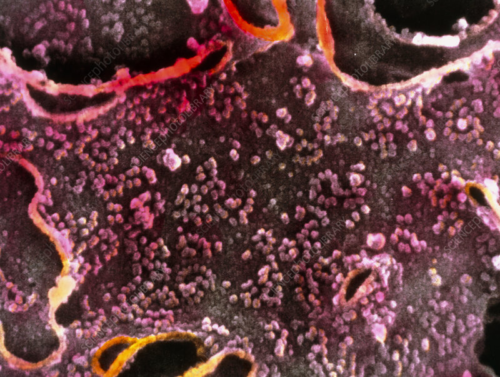
The plethora of proteins within us are produced by a cellular structure that, if not for its intricate folding, would be ten to twenty times larger than the outer surface of the cell itself. Its name is the endoplasmic reticulum (ER), and it serves as the production facility where proteins are assembled, folded, and secreted. Seeing how crucial proteins are in maintaining metabolism, it is no surprise that the ER works very hard to keep us alive.
Unfortunately, things don’t always run smoothly: the ER sometimes over-exerts itself. If you get sick, for instance, each day, the mature B-cells of your immune system will secrete up to their own weight in antibodies, which are all processed in the cells’ ERs. Glucose deprivation and calcium regulation also affect processes in the ER by triggering responses that interfere with protein folding. The imbalance between supply and demand of properly shaped proteins produced by the ER leads to a phenomenon called ER stress, which may lead to type 2 diabetes, atherosclerosis, and liver disease.
Fortunately, our cells have a set of proteins that sense ER stress and respond to stop it. These proteins, collectively called unfolded protein response (UPR) proteins, play a major role in cellular life and death decisions—this feature has garnered an intense interest in the role of UPR proteins in many diseases. However, there is a lot we do not know about these proteins and their roles.
The most common of these proteins is IRE1α. Initially, IRE1α calls for molecular chaperones that guide misfolded structures along the proper pathways for folding, helping the ER return to its normal functioning conformation. However, if activated for too long, IRE1α starts to initiate apoptosis, or programmed cell death, through the signal-regulating kinase 1 (ASK1) protein and through proteins called caspases. It makes sense that activity is tightly regulated, but how this happens is unclear. Unveiling the molecular mechanisms that govern IRE1α activity could help us understand how UPR proteins are regulated across all eukaryotic cells, as well as how a number of human diseases develop.
Research conducted by Malaiyalam Mariappan and Xia Li at the Yale School of Medicine has helped shed light on this intricate process. Their work describes how UPR proteins are regulated. “We identified the mechanism that actually turns off the IRE1α sensor molecule so that cells can get back to normal, rather than going into cell death,” Mariappan said.
The key, they found, was a protein called sec63, which actively recruits a protein chaperone called binding immunoglobulin protein (BIP) to stick to IRE1α like a cap. This capping prevents IRE1α from binding to itself and forming long, active, peptide chains. Thus, it forces IRE1α to exist in an inactive state as singular proteins.
Under normal conditions, there is plenty of BIP for sec63 to pick up. During ER stress, BIP preferentially sticks to misfolded proteins instead of IRE1α, and this lack of capping allows IRE1α to bind to itself and activate. Active IRE1α chains initiate cell transcription pathways that produce more BIP, and it is this self-regulating negative feedback mechanism that keeps IRE1α in check.
The discovery of sec63 as an inhibitor of IRE1α clustering is an addition to the Mariappan lab’s long line of important findings on regulators of proteins such as IRE1α. “Initially it was thought that IRE1α was an independent protein that sensed ER stress and sent signals to the nucleus… but we were the first to report that it’s actually part of a big complex with protein translocation channels,” Mariappan said.
Elucidating the role of sec63 as a recruiter for BIP opens up a host of new questions. “Still in this field, there’s a lot of mystery, especially in ER stress and the unfolded protein response field,” Li said. “I just want to know how it all works.”
Citations
Carter, C. S. (2017). The Oxytocin–Vasopressin Pathway in the Context of Love and Fear. Frontiers in Endocrinology, 8. doi:10.3389/fendo.2017.00356
Katsube, H., Hinami, Y., Yamazoe, T., & Inoue, Y. H. (2019). Endoplasmic reticulum stress-induced cellular dysfunction and cell death in insulin-producing cells results in diabetes-like phenotypes in Drosophila. Biology Open, bio.046524. doi:10.1242/bio.046524
Li, X., Sun, S., Appathurai, S., Sundaram, A., Plumb, R., & Mariappan, M. (2020). A molecular mechanism for turning off IRE1α signaling during endoplasmic reticulum stress. Cell Reports, 13(33). doi:10.1101/2020.04.03.024356
Lin, J. H., Walter, P., & Yen, T. S. B. (2008). Endoplasmic Reticulum Stress in Disease Pathogenesis. Annual Review of Pathology: Mechanisms of Disease, 3(1), 399–425. doi:10.1146/annurev.pathmechdis.3.121806.1514
Milo, R., & Phillips, R. (2016). Cell biology by the numbers. New York, NY: Garland Science, Taylor & Francis Group.
Ponomarenko, E. A., Poverennaya, E. V., Ilgisonis, E. V., Pyatnitskiy, M. A., Kopylov, A. T., Zgoda, V. G., … Archakov, A. I. (2016). The Size of the Human Proteome: The Width and Depth. International Journal of Analytical Chemistry, 2016, 1–6. doi:10.1155/2016/7436849
Xu, C. (2005). Endoplasmic reticulum stress: cell life and death decisions. Journal of Clinical Investigation, 115(10), 2656–2664. doi:10.1172/jci26373

