Image courtesy of Flickr.
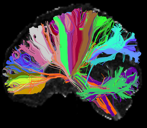
For decades, researchers have sought to map the “connectome,” a structural wiring diagram of brain communication. Yale School of Medicine Professor of Radiology and Biomedical Imaging and of Neurosurgery and Director of Magnetic Resonance Imaging (MRI) Research Todd Constable and his lab are more interested in the functional connectome—the organization of neurons working together during different tasks.
Blood flow and neuronal activity are correlated, allowing active brain regions to be mapped via functional MRI, which assembles images made of 3D volume-pixels (“voxels”) using blood-oxygen-level-dependent signals. Atlases (coordinate maps of brain structures describing outline or volume) are constructed by parcellating regions into fewer hubs called nodes based on functional synchronization, but there’s no consensus on a singular atlas, making it difficult to compare and generalize findings that aren’t dependent on the same atlas.
“When there’s dysfunction in certain circuits, you want to be able to point and talk about them in a reasonable manner where people know what it is you’re talking about. And right now, we don’t really have that common language,” Constable said.
Finding an ideal, stable atlas was the challenge Wenjing (Wendy) Luo, a Ph.D. student in the Yale Department of Biomedical Engineering, and Constable originally sought to tackle. However, they discovered something many studies have long taken for granted.
The functional connectome is a matrix of nodes. Past research examined between-node changes and connectivity strength, but a study demonstrating changes in nodes across tasks inspired Luo and Constable’s recent study published in NeuroImage examining within-node changes. “We were trying to find out what is actually driving node reconfiguration, and that led us to look at smaller-scale connectivity,” Luo explained.
Using machine learning to analyze data from the Human Connectome Project S900, Luo’s models displayed strikingly high accuracy in matching subjects’ scans in one task (e.g., resting, gambling) to their corresponding scan in a different task, predicting the task performed in scans, and classifying subject gender. Additionally, by comparing singular voxels to others in the same node, she found that differences in inter-voxel connectivity affect node connectivity as a whole. The shift in voxel connectivity across tasks and conditions implies that neural activity within nodes is far from uniform and unchanging, as had been previously assumed.
Ignoring within-node changes means loss of crucial information that could improve models’ predictive power of a person’s attention and memory and associations of behavior with brain activity. “If we take this information into account, lots of results of some previous studies should change,” Luo reflects, referencing her paper which identified 50 studies that potentially misattributed results to between-region changes rather than changes within regions themselves. “So we have to be careful about many claims that have been made before.”
Constable has another important takeaway. “The brain is complicated. In the neuroimaging community, people keep wanting to boil things down to this region does this or this region does that. And I think that’s the wrong approach. There are sophisticated systems that are flexible in how they team up to execute functions, and we need to embrace their complexity.”

