
How we physically feel things—and how we form perceptions of the world based on what we feel—is an age-old question in the field of neuroscience. Penetrating into the different layers of our skin lies a network of tiny sensory structures called mechanoreceptors. These specialized structures respond to tactile stimuli—everything from the pressure of a handshake to the tickle of a breeze—converting touch into electrical signals that travel to the brain. When something presses against the skin, it creates tiny deformations in the underlying tissue, activating these mechanoreceptors to produce electrical signals known as action potentials. These electrical signals travel along primary sensory neurons to the spinal cord and are eventually relayed to the somatosensory cortex of the brain, a region where tactile information is processed.
As it turns out, we currently have a limited understanding of one of these mechanoreceptors, known as Pacinian corpuscles. Reevaluating the current model of the Pacinian corpuscle could provide critical insights into the biological pathways responsible for touch and sensation.
Yale neuroscientists Luke Ziolkowski GSAS ’25 and Yury Nikolaev, along with Slav Bagriantsev, a professor of cellular and molecular physiology at the Yale School of Medicine, recently investigated the structure and function of the Pacinian corpuscle. “You have to go back to papers from the 1950s and ’60s to find good research that looked into the functional mechanisms of how Pacinian corpuscles work,” Ziolkowski said. Today, researchers have far more advanced tools to investigate both the structure and function of Pacinian corpuscles, allowing for deeper insights than were possible before. The deeper insights are revising some prior findings. While some kinds of mechanoreceptors constantly signal the presence of a stimulus, the Pacinian corpuscle is part of a mechanoreceptor group that responds only to transient stimuli. Action potentials are only fired from the corpuscle when a stimulus is first applied and again when it is removed; this pattern of neuronal firing is known as rapid adaptation.
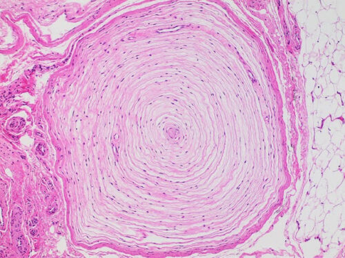
The Pacinian corpuscle consists of onion-like layers surrounding an inner core. Within the inner core is the tip of a sensory nerve, known as the afferent terminal, which sends signals to the brain. According to the classical model, the multilayered outer core acted as a mechanical filter for touch, blocking steady pressure or slow vibrations so that only rapid changes in touch reached the nerve terminal. Furthermore, it had been assumed that only the nerve terminal itself was capable of detecting tactile stimuli. Through a combination of structural and functional studies, the researchers were able to successfully challenge both of these long-standing hypotheses and propose an updated model of how the Pacinian corpuscle functions.
To revise the hypothesis, the researchers started by studying the bill skin of a late-stage embryonic mallard duck to determine the architecture of the Pacinian corpuscle. They used data from a high-resolution microscope to reconstruct the corpuscle in 3D, which allowed them to visualize the interactions between its components. The 3D model revealed that the inner core is physically connected to the nerve terminal, suggesting that the two structures may interact much more closely than previously thought.
Next, the researchers assessed whether the outer core is necessary for sensing touch. Using Pacinian corpuscles from the bill skin, the researchers ruptured the outer cores, decapsulating the corpuscles, and directly stimulated the inner cores with vibrations of various frequencies. To assess the nerve’s response, they measured the tiny electrical currents in the nerve terminal. The nerve terminal was found to respond most strongly at the start and end of the vibrations, as well as at higher frequencies, demonstrating the retention of the rapid adaptation process. Nonetheless, the outer core is still needed to create a favorable environment for the inner core. “Once you remove the outer core, the inner part that senses mechanical force can still function, but only for a short period of time,” Bagriantsev said. The experiment displayed that the outer core, while still necessary for the integrity of the inner core, is not directly involved in sensing transient touch.
Finally, the researchers turned to the inner core to investigate the origin of function in the Pacinian corpuscle. They looked at a type of glial cell found in the inner core called the lamellar Schwann cell (LSC). The researchers stimulated the LSCs and found that they produced action potentials. The nerve terminals, in turn, then generate action potentials, indicating that the LSCs of the inner core contribute to the sensitivity of the nerve terminal. “LSCs act as amplifiers of mechanical stimuli, helping the neuron detect touch,” Bagriantsev said. While 3D modeling has displayed the physical connection between LSCs and the nerve terminal, this experiment provides evidence of a functional connection as well. “A non-neuronal cell contributing to touch detection is a whole area that people didn’t really think about before,” Ziolkowski said. The researchers were able to prove that the nerve terminal, in opposition to previous beliefs, is not the only component of the Pacinian corpuscle where physical sensation is perceived.
How we feel things—in particular, transient, high-frequency vibrational stimuli— is dictated by the microscopic onions beneath our skin whose detailed description has led to a revised model. The outer core of the Pacinian corpuscle, thought to serve as a mechanical filter, is dispensable and appears to primarily play a protective role over the inner core, whose LSCs perceive tactile sensation in addition to the afferent nerve terminal. “The part we don’t know yet is how information is being communicated from the LSCs to the neuron itself,” Ziolkowski said. “That mechanism is something that is continuing to be looked at.”
The implications of this research are enormous, particularly in the field of touch-based technologies. “There is a demand for coming up with mechanical sensors for different applications, including next-generation prosthetics,” Bagriantsev said. “There are a lot of efforts that learn from mechanoreceptors and are trying to build physical sensors based on what we know from biology.” By revisiting a decades-old scientific paradigm, Ziolkowski, Nikolaev, Bagriantsev, and their team were able to form a new model for the Pacinian corpuscle that may soon inform the sensory technologies of the future.