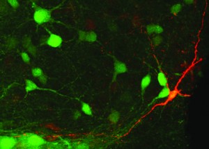
Research by professor Christopher Pittenger at the Yale School of Medicine has found that interneuron destruction in mice promotes Tourette’s-like symptoms. This finding is a key step in unlocking the poorly-understood mechanisms of Tourette’s Syndrome, a condition that causes uncontrollable, repeated movements in patients, especially when under stress.
Pittenger decided to focus his work on interneurons when he compared brain tissues of mice with Tourette’s to brain tissues of wild-type mice — he observed fewer interneurons in the rodents with Tourette’s. Still, he did not know whether the reduced interneuron population resulted from disease or from a consequence of treatment. His recently published work set out to answer this question.
Pittenger wanted to test whether loss of neurons directly leads to reduced neural function. In order to do this, he developed a method that uses diphtheria bacteria to destroy motor neurons in the brain. Pittenger noted that the most difficult part of the study was developing this destruction method. It required a combination of procedures: He needed to induce the expression of diphtheria receptors in interneurons, and he needed to control the incorporation of receptors to target specific interneurons.
The study revealed that interneuron loss leads to excessive grooming in mice that have experienced stress. Since this behavior is consistent with Tourette’s, Pittenger concluded that interneuron removal is indeed linked to the emergence of Tourette’s tics in mice. “This conclusion represents a large step in unraveling both the complex neural mechanisms behind disease, as well as an insight into the role of interneurons in mediating mental health,” Pittenger said.
