You lost your keys again. Without any idea as to where they went, you dart from room to room, pursuing each lead for just a moment before giving up and trying something else. As you search, you piece it together and suddenly remember where they are: the refrigerator. Turning away from your hamper, you make a beeline for the kitchen, and the keys are recovered within moments. The way you went about finding your keys—the indiscriminate giving way to the direct—is not unique to forgetful humans. All living things – dogs, bees, cancer cells – devise these strategies as they explore the world around them. In the last case, however, this probing can do more harm than good. The more cancer cells move in a persistent beeline, the more efficiently they will spread and the more lethal the cancer will be.
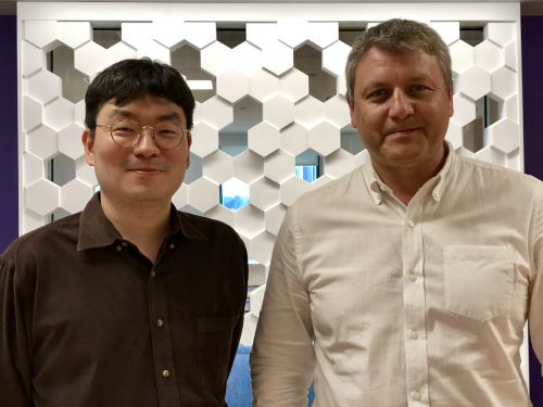
A team at the Yale Systems Biology Institute led by Professors Andre Levchenko and postdoctorate researcher JinSeok Park wanted to more comprehensively understand how cancer cells find their proverbial keys. To start, it is important to understand how cells interact with their direct outside environment, a network of proteins called the extracellular matrix (ECM) that cells secrete themselves. Coming into contact with a neighboring cell’s ECM triggers two signaling pathways that mediate cell movement. The balance between the different behaviors that these two pathways trigger—the cell’s polarity, so to speak—influences cells’ migration behavior, their switch from frenzy to beeline. Previous research implied that cell contact with the ECM was responsible for these different patterns, but how the cells went about this was poorly understood. Levchenko and Park derived a mathematical model to predict a given cell’s behavior based on the activity of the two principal signaling pathways, which both cements the link between cell-ECM contact and migration and, more importantly, clarifies a new avenue for cancer treatment.

Migration
The type of cancer called melanoma rarely kills at its origin, the skin, but rather takes lives when it attacks other organ systems, or metastasizes. Therefore, it is a disease that relies heavily on movement, a trait that made it a useful model for the team to study. The critical moment in melanoma progression happens when the cancer stops expanding across the skin and begins tunneling downward. At that point, it is only a matter of time until melanoma cells find their way to a blood or lymphatic vessel, the anatomical highways necessary for metastasis. But first, these cells have to navigate the fibers of the dermis, the layer of tissue right below the skin.
These tissue fibers are like ropes that cells can pull to move around. There are three types of cell movements. When the cells rapidly pull ropes in different directions, their behavior is random; when cells alternate pulling forward and backward on the same rope, their behavior is oscillatory; and when cells consistently pull one rope in one direction, their behavior is persistent. The oscillatory pattern is a cell-specific behavior and complicates the model—once a target has been established for the cell, it continues to travel back and forth in the vicinity instead of heading straight there. Levchenko likes to think of oscillation as a cell overshooting its target and doubling back repeatedly. Regardless, if a cell is to reach a destination, persistent behavior is the most efficient, which, in the context of cancer, means a faster progression towards metastasis.
Migration patterns do not happen by chance. Cells’ direct contact with the fibers of the ECM sets off two sets of signals that influence cell movement and so determine whether cells will display random, oscillatory, or persistent behavior. One of these signals is characterized by a protein called Rac1 and the other by a protein called RhoA. The two sets of signals work towards the same goal—to mediate cell movement—but perform opposite functions, as Rac1 acts as an accelerator to RhoA’s brakes. To complicate matters further RhoA functions to halt cell migration, its presence is also required for migration in the first place. This somewhat paradoxical mechanism only highlights the complexity of these networks. Research in oncology typically has focused on how cancer cells grow and replicate, but these convoluted and poorly understood processes of cell migration pushed Levchenko and Park to establish a coherent model and reconcile the three migration patterns.
Polarization
The model the researchers derives uses advanced mathematics to integrate these three behaviors into the same framework, an unprecedented feat. The model shows how cells’ contact with the ECM changes the balance between the levels of activity of the Rac1 and RhoA pathways, which in turn impacts migration. By plotting the two activation rates on the same axes, the researchers were able to establish discrete regions for each migration pattern. For example, if a cell has a high Rac1 activation rate and a low RhoA activation rate—a lot of accelerator and little brakes—it will display persistent behavior.
The model accounts for not only the balance, but also the location of this pathway activation within a given cell. By tracking sites of high signaling activity, the researchers were able to visualize the spatial distribution of these two pathways. In randomly migrating cells, signaling was sporadic; in oscillating cells, hotspots of activity alternated between the front and the back; and in persistently moving cells, activity was high on one side and inhibited on the other. These signals are consistent with the aforementioned “ropes” of the ECM and helped Levchenko and Park affirm that their model accurately represented cell movement.
This model is unique because Levchenko and Park were able to map specific populations of cells onto diagrams that were generated using the model. Within the same cancer or even the same tumor, different cells will display different behaviors, and the model accounts for these varied responses. “[Cells] all get the same piece of information, but they interpret that information in very different ways,” said Levchenko. Understanding how real cells move within and correspond to the rigid mathematical model was an especially rigorous step that confirmed the model’s validity.
Manipulation
Once a model had been established, the next step was to manipulate it. One determinant of cell migration is the density of the ECM. To mimic the ECM, the researchers placed artificial nanoscale posts on a cell adhesion surface. As more posts were added, the fraction of persistently migrating cells decreased. “[The cells] are getting too much input from the surface, so they are confused,” Park said. When there were fewer posts, cells seemed to perceive their organization into rows and so had stronger directional cues, but when there were many posts, the cells were less able to navigate. The proportions of the migration patterns as predicted by the model were backed up with experimental data from real melanoma cells, and as expected, the more persistent cell migration was, the farther cells migrated.
Levchenko and Park went on to alter a whole host of factors in order to understand the model’s viability outside their tightly regulated conditions. When either the Rac1 or RhoA signaling pathway was perturbed, the fraction of persistently moving cells decreased, a finding that is consistent with the networks’ modulation of cell movement. Similarly, when a drug that inhibits the formation of microtubules, elements of the cytoskeleton that play a role in migration, was administered, persistent cells decreased. Lastly, the researchers tested whether using a different cell line would yield the same results. All the previous experiments were done in a cancer cell known to be particularly invasive. To determine whether the model would fit a less aggressive melanoma, it was tested in a non-invasive cell line for which fewer persistent cells were expected, as a less invasive cancer metastasizes less efficiently. Despite the change in the aggressiveness of the cancer, the model held, which suggests its application in a much broader context of cancer therapy.
Humanization
To expand their discovery, Levchenko and Park hope to shed further light on the role of cell-ECM communication in cancer research. Once a complete map of the key signaling networks that dictate a cancer’s behavior is understood, targeted therapies can infiltrate them. If the networks can be manipulated to limit the proportion of persistently migrating cells, then even a cancer that has begun to spread can be delayed. Prolonging the period of time before a cell makes a beeline for a blood or lymphatic vessel in this fashion is an untapped therapeutic avenue towards which this model makes great strides. On the clinical side, the model allows for much more accurate predictions of the interval of time between the onset of a primary tumor and the cancer’s metastasis, which can undoubtedly improve cancer prognosis.
Cancer treatment is ultimately about patients’ well-being. If this model can delay the cell search and prevent persistent cell migration, it could prolong life by months, which can make a significant difference for a highly aggressive cancer like melanoma, which has only a 15-20% 5-year survival rate in its most advanced form. Understanding how we can target the molecules that modulate cells’ search period is an important therapeutic step towards an increase in these rates. Maybe one day melanoma cells will never find their keys.
About the Author
Charlie Musoff is a sophomore in Davenport College and a molecular, cellular, and developmental biology major. Besides being Yale Scientific’s Outreach Designer, Charlie enjoys running, singing with the Baker’s Dozen, and teaching with Community Health Educators.
The author would like to thank Professors Andre Levchenko and JinSeok Park for their time and insights.
Further Reading
Holmes, William R., et al. “A Mathematical Model Coupling Polarity Signaling to Cell Adhesion Explains Diverse Cell Migration Patterns.” PLOS Computational Biology, vol. 13, no. 5, 4 May 2017, doi:10.1371/journal.pcbi.1005524.

