
Cells are notoriously chatty. Communication between different cell and tissue types is happening constantly in any multicellular organism, and this crosstalk is critical to survival. In humans, chemical signaling pathways ensure that information in one tissue can be shared with others, linking the whole body together as one growing, changing organism. While biologists largely focus on the role of proteins in cell-to-cell communication, a team of chemists discovered the importance of small molecule crosstalk in ovarian cancer.
Researchers in Laura Sanchez’s lab at the Department of Medicinal Chemistry and Pharmacognosy at the University of Illinois at Chicago recently developed a method to analyze the spatial and temporal changes of a chemical landscape in a cell culture—to see cells talking. Rather than looking at changes in protein expression within “sending” and “receiving” cells, this new method captures a snapshot of what’s happening with small molecules outside the cells by culturing different cell types together and seeing how the molecules present in that culture change over real time.
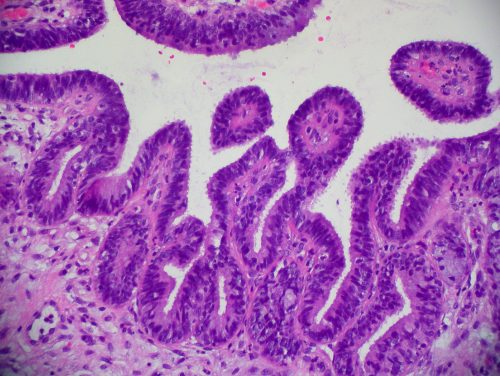
Understanding cell signaling pathways is especially important when the communication goes wrong. In this study, the researchers investigated the role of cellular communication in high grade serous ovarian cancer (HGSOC), a highly lethal gynecological cancer that cannot be detected early and whose mechanisms are poorly understood. Recent findings that HGSOC tumors actually originate in the nearby fallopian tube, not the ovary, raised the question of whether chemical signaling could be crucial to this early metastasis, or the movement of cancerous cells from their native tissue to new tissues.
Underlying this new method is one of the most common yet powerful tools in chemistry, mass spectrometry (MS). MS identifies molecules in a sample by determining their mass to charge ratios. Imaging mass spectrometry (IMS) uses optical tools to add a spatial component. “Normally you’re doing an extraction and losing spatial information. Imaging mass spec is powerful because you can make these maps of chemistry across a sample,” said Sanchez.
The study modeled HGSOC cellular interactions by co-culturing mouse ovaries with cancerous fallopian tube cells in agarose. “People had been looking at tissue slices for a long time, but [that method is] a static time point, [and] you need to have a diseased condition versus a healthy condition,…so we needed to develop something where we could incubate and allow for exchange to happen on the same platform that we imaged,” said researcher Katherine Zink, a graduate student in the Sanchez Lab. Some crucial elements of this method included running all control and experimental samples on the same 8-well plate and confirming that drying the agarose—which captures the chemistry in 2D for IMS analysis—did not stress or kill the cells.
After optimizing their culture, the researchers performed IMS and identified a mass/charge peak that significantly increased with the presence of both ovary and cancerous fallopian tube cells. By looking through the literature for previous identification of similar peaks or small molecules implicated in HGSOC, the researchers eventually found an exciting hit: the small molecule hormone norepinephrine. Among other research, a 2015 study found that the median survival rate for ovarian cancer patients who were on medications that block norepinephrine signaling was greater than other patients by almost 60 months. For Sanchez, it was exciting to find such immediate relevance to their work. “What we’re finding in the lab can be directly translated to human outcomes,” she said. The researchers then validated the peak assignment with other chemical techniques to remove any doubt that norepinephrine was the molecule they were seeing. “To be compliant with good chemical practices, we really wanted to have something other than mass to confirm that [norepinephrine] was the molecule…I think it’s very definitive now that there’s no other way you could make this [mass/charge peak] assignment,” said Sanchez.
The discovery of increased norepinephrine in ovary/fallopian cancer cell co-cultures not only validated the biological evidence of norepinephrine’s role in ovarian cancer to guide future research into this cancer’s mechanisms, but it also answered the more fundamental question of whether there is any chemical crosstalk in cancer. The very fact of chemical signaling between cancerous and noncancerous cells is a new discovery, noted Sanchez, as cancer research usually focuses on enzyme pathways.
Importantly, the applications of the optimized culturing and IMS methods can answer questions about the chemistry underlying all sorts of coordinated processes all over the body—with other cancer types, for example, or even in the brain. Sanchez says it’s just a matter of plugging different cell types into this system. As she put it, “The sky’s the limit if you can grow it in agarose”.