
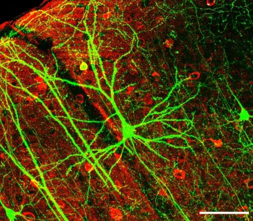
There’s an old saying that “a common man marvels at uncommon things, while a wise man marvels at the commonplace.” For both the common and the wise man, nothing is more commonplace nor more worthy of marvel than one’s own brain. Every second of every day, billions of brain cells called neurons communicate with each other using electrochemical signals, working together to create awesome cognitive processes, behaviors, emotions, and memories. A symphony of consciousness is constantly playing between your ears, each crescendo of neural activity making up the world as you experience it, from how you think to the things you see, to the movement of your body. Phenomena like these are the products of complex, finely-tuned neural mechanisms and circuits. Despite the wide array of tasks our brains perform, the building blocks of communication between neurons throughout the nervous system is universal.
Curious clustering
Synaptic transmission is the means by which neurons communicate with each other and with other tissues in the body. Each neuron, once triggered by a sufficiently strong electrical signal, will in turn fire another signal down its long, cable-like part of the cell body called an axon. This signal will propagate down the axon until it reaches the branched end of the axon, or axon terminal. In the body, the terminals of different neurons sit very close to each other—separated by small gaps called synapses. To send a signal across the synapse, a neuron must release chemical messengers called neurotransmitters. The neuron packages the neurotransmitters in little bubbles of membrane called vesicles, which sense electrical signals arriving at the axon terminal and releases its neurotransmitter contents into the synapse. The neurotransmitters are then received by adjacent neurons, which initiate a signal down its axon, starting the process over again.
This method of intercellular communication is fascinating as a whole, but prompts questions at each step of the way. One such question, which caught the eyes of a Yale research team led by Pietro De Camilli, pertains to the clustering of synaptic vesicles at the surface of axon terminals. When synaptic vesicles reach the target terminal, they coalesce into tightly-packed clusters of hundreds or evens thousands of vesicles. These vesicles, despite being tightly packed together, very quickly diffuse and release their contents upon an activation signal from the pre-synaptic terminal. “And so, the question that arises is: how are these vesicles kept together in a cluster while still maintaining their high mobility?” said Dragomir Milovanovic, a postdoctoral fellow working in the De Camilli lab.
Liquid-liquid phase boundaries
The first clue toward the mechanism behind clustering came from the vesicles’ fluid-like behavior. When the newly formed vesicles are chemically labeled and the clusters at axons are observed, the researchers found that the freshly-formed vesicles are mixed evenly with the unlabeled vesicles at the nerve terminal, rather than sticking onto the edge of these preexisting clusters. This mixing behavior stands in stark contrast to the very tight, sharp boundary that the clusters have overall. “How could a crosslinking matrix be compatible with such fluidity? One could not see a retaining membrane or matrix around synaptic vesicles, yet, synaptic vesicle clusters had sharp boundaries,” said De Camilli. A recent flurry of papers demonstrating that different proteins are able to separate from the cytosol into a distinct liquid phase led the De Camilli lab to hypothesize that perhaps the clustering of synaptic vesicles could be explained by the properties of liquid-liquid phase boundaries.
A liquid-liquid phase boundary occurs when a protein in solution is able to weakly link up with other proteins. The result is a little bubble of the protein, with distinct boundaries keeping it from mixing in solution. If you’ve ever mixed oil and vinegar, you’ll have witnessed a similar phenomenon. Like oil droplets floating in vinegar, many proteins, such as RNA, form these bubbles in intra- and extra-cellular fluid, performing important biological functions. With the biological importance of liquid-liquid phase interactions in mind, the De Camilli lab set out to discover whether synaptic vesicle clustering could be explained by a liquid-liquid phase boundary model, rather than a model describing the vesicles as rigid structures made of scaffolding proteins.
Synapsin: the missing link
For a liquid-liquid phase boundary to occur, there needs to be a special type of protein in high abundance that is able to engage in multivalent interactions—that is, it needs to be able to stick to more than one protein at a time. It also needs to have a relatively weak affinity for all proteins so that it can readily diffuse away when necessary.
The De Camilli lab found, after some searching, a viable protein that met the parameters for liquid-liquid phase separation: synapsin. Synapsin is highly abundant in the neuron, and is known to possess a large intrinsically disordered region (IDR), which is an area of a protein that is unable to fold into a stable configuration. “Since it has been suggested that these proteins with these IDRs sometimes can phase separate by themselves, I decided to look at the synapsin alone,” said Milovanovic.
Indeed, when placed into solution by itself, synapsin formed droplets with liquid-liquid phase boundary characteristics; the synapsin droplets have an affinity for each other, coalescing into larger droplets or clustering with other droplets around it. Additionally, when the synapsin in one of these droplets is tagged with a fluorescent dye and then photobleached with a strong laser to deactivate the dye, the droplet recovered its fluorescence over time—evidence that the clusters are indeed acting as a liquid, exchanging photobleached synapsin with the still-fluorescent synapsin in other clusters. A solid vesicle structure with rigid scaffolding proteins would be unable to regain fluorescence, but a droplet with a liquid-liquid phase boundary is able to mix with other droplets around it, swapping out bleached synapsin for unbleached synapsin.
With the liquid-liquid phase boundary nature of synapsin established, the lipid sequestering capability of synapsin clusters was then tested. Lipids are biomolecules that make up the membranes of vesicles, including synaptic vesicles. If synapsin plays a role in vesicle clustering, it should be able to sequester lipids. Lipid vesicles similar in composition to those of a synaptic vesicle were prepared and then mixed with synapsin. After some time, the synapsin once again formed droplets, but this time they were enriched with the lipid vesicles, altogether forming clusters of synaptic vesicles that closely resembled the clusters normally found at pre-synaptic terminals. These results demonstrate the liquid-liquid phase boundary nature of synapsin and its ability to cluster synaptic vesicles in the same way as observed in nature.
Synapsin’s ability to form these clusters was established, but further study was required in order to determine how critical synapsin’s role was in vivo. Therefore, the team employed the existing model of synapsin triple knockout mice in which three genes responsible for producing synapsin were deleted. They looked at the nerve terminals of brain sections from synapsin triple knockout mice and compared these nerve terminals with the ones of wild-type mice. They found that the mice without synapsin had less than fifty percent fewer synaptic vesicles present at synaptic terminals than wild-type mice, and the vesicles that were present had very limited clustering capabilities.
The team had one last task: to explain the rapid mobility of the vesicle clusters. In vivo, when a neuron depolarizes and sends a signal down its axon, the axon terminals are flooded with various enzymes including CaMKII, which is known to play a role in vesicle release into the synaptic cleft. The researchers found that when synapsin clusters are exposed to CaMKII, the droplets dispersed very rapidly, mimicking the in vivo behavior of vesicle clusters when a signal arrives at the axon terminal.
Far-reaching implications
Overall, the team seems to have found an explanation for the behavior of vesicles. What confused previous researchers—the paradoxically rigid-yet-fluid clustering, combined with the high mobility of vesicles—was elucidated by researchers in De Camilli lab, who discovered the connection between liquid-liquid phase boundaries and synapsin function.
This discovery may also prove valuable to other researchers working both in and outside the realm of the neuron. Prior to their work, all examples of liquid-liquid phases in literature referred only to those involving proteins, RNA, or protein and RNA mixtures. “Ours is the first clear example of a cytosolic liquid phase that comprises vesicular organelles. I think that other vesicular compartments in the cells may assemble via the same principles,” concluded De Camilli.