
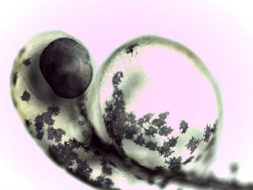
Every one of us started out as an undifferentiated mass of cells known as the embryo. This small cluster of cells is not unlike a busy city at rush hour, with thousands of cells buzzing about, the hustle and bustle coordinated by orders in the form of chemical signals. These signals determine each cell’s destination. Some cells stay roughly where they are, but most cells undertake grand migrations to various poles of the embryo where they undergo differentiation, ultimately adopting their designated vocation.
Trying to visualize this intricate dance can be an awe-inspiring experience. For some researchers, the ultimate goal is to fully understand the complex network of mechanisms that bring a messy collection of cells into a highly organized being. The Holley Lab in the Department of Molecular, Cellular, and Developmental Biology attempts to piece apart these mechanisms using zebrafish embryos.
It was previously known that a part of the vertebrate embryo known as the tail organizer is a vital information center, which creates and distributes chemical signals that determine the fate of various cells. While studying these processes, researchers in the Holley Lab found for the first time that the tail organizer’s influence extends beyond the range of its chemicals by relaying mechanical information. Knowledge about the transmission of mechanical information offers insight into the nature of cell mediums.
Throwing the stone into the water
The discovery of mechanical force as a means of organizing cells was a fortuitous one. It came while the researchers were studying the role of the tail organizer as a key player of embryonic development. “What we thought we were going to do was perturb this organizer and see exactly how it regulates morphogenesis—the surprising thing is this mechanical information that came out of it,” Holley said. In developmental biology, an organizer is a region of cells with the ability to determine the fates of surrounding cells and steer their journey into becoming specific types of tissues. In an embryo, the tail organizer secretes molecules known as morphogens, thereby establishing a chemical gradient. Cells in regions of different morphogen concentrations are then directed to different cell fates. An important class of morphogen is bone morphogenetic proteins, or BMP. BMP is a key chemical signaler that facilitates body elongation in embryos and coordinates cell migration.
The story of this work thus began with the team’s attempts to study the role of the tail organizer. This research was conducted by deforming zebrafish embryo and modifying the amount and pattern of BMP secretion. Emilie Guillon, a postdoctoral researcher involved in the project, likens their approach to throwing a stone in water to observe the physical outcome. “To know the speed of sound in water, you throw a stone in water to see how fast you are propagating that sound, and one way to prove the properties [of tissues] is to create a perturbation,” Guillon said.
The researchers perturbated the system in two ways. They treated the embryos with DMH1, a chemical that nullifies BMP activity in the tail organizer, and concurrently overexpressed eve1, a gene known to be involved in embryonic development. While DMH1 causes abnormalities on its own, the most severe defects in embryos were observed in a previous study when a combination of chemical and genetic perturbations were applied. The eve1 gene is a member of a gene family central to establishing the body plan of an embryo and plays an important role in embryonic elongation. This procedure demonstrates a general approach in biological studies: to learn about the importance of a molecule or gene, the quickest route is often to tinker with its function. By observing what goes wrong when the component in question is not functioning properly, one gains insight into the normal role that component plays in a system.
Reading the ripples
With chemical and genetic tools to understand embryonic development in hand, the team observed which specific locations of the embryo were impacted by agitating the system. To this end, the researchers employed fluorescence microscopy to target and illuminate the BMP cell surface receptors when they are bound by BMP, thereby allowing the scientists to visualize the distribution and activity of BMP in the cell. They found that fluorescence appeared only in the tail organizer, suggesting that BMP signaling did not extend throughout the rest of the embryo. The team surmised that irregular cell behavior would be found exclusively in the tail organizer where BMP was located and set out to look for it.
Irregular cell migration was detected not only in the tail organizer, but also in the posterior neural tube, a region of the embryo where cells are expected to travel in a streamlined manner. This mishap in cell motion contributed to the abnormal embryos the team observed, as precise patterns of cell migration are important for proper embryonic development. Crucially, the posterior neural tube is outside the reaches of BMP signaling—had BMP been the sole mechanism using which cells were organized, there could not be abnormal cell migration elsewhere. This finding came as a surprise to the researchers, who then postulated that mechanical force could be involved in transmitting information and organizing the cells.
Turning to simulations
To test their hypothesis that mechanical information played a role in development, the team turned to Thierry Emonet, professor of Physics and Molecular, Cellular, and Developmental Biology at Yale, as well as to Corey O’Hearn and Mark Shattuck, professors of Mechanical Engineering and Physics, respectively. Emonet, O’Hearn, and Shattuck created a computer model to simulate the behavior of cells under mechanical perturbations, providing predictions of cell behavior that the researchers could then attempt to find in the real embryos. In the model, cells were represented as spherical particles, and a disturbance was initiated by increasing the repulsive force from cells in the tail organizer—reflecting the perturbation the team had previously caused in real embryos. In the model, mechanical perturbation led to an increased probability of backwards motion in the modeled cells. The team also found that cells in the model had an increased density in the posterior neural tube in a manner consistent with the jamming of cells—not unlike a traffic jam. Here, the jam starts at the accident site, and then propagates backwards as a retarding force away from the site, spreading the disturbance. This would be consistent with the simulated cells transferring mechanical information in the same manner as waves that are transmitted through bulk material—that is, in the form of compressions. “It is like a kind of a wave, like in the ocean, that is travelling through the posterior neural tube,” Guillon said.
To determine if the findings of the computer model on cell motion mirrored those in actual embryos, the team looked at real zebrafish embryos to see if similar backwards cell motion occurs when the cells are perturbed with DMH1 and eve1. The researchers observed that cells did, in fact, move abnormally backwards, mirroring the simulation results. Moreover, they also observed a widespread decrease in the coherence of cell motions, suggesting that the hypothesis of mechanical information transmission is correct.
Plugging the holes
To further support their case, the team considered the alternate hypothesis that cells abnormally move outside the range of BMP signaling due to a mishap in chemical signaling caused by a still unknown mechanism of concerted action by DMH1 and eve1. It was possible that other morphogens important for body elongation, such as Wnt1 or Fgf, could be impacted upon the addition of DMH1, thereby having the same effects as mechanical perturbation. “What if actually the abnormalities were still chemical—caused by actually perturbing Wnt and Fgf pathways?” Guillon said. To eliminate this possibility, the team studied the Wnt1 and Fgf signaling pathways in the perturbed embryos. They found that the addition of DMH1 did not impact these signaling pathways. At this point, the team also found that that the abnormally backwards-moving cells travelled at speeds much faster than chemical information could travel, strengthening the mechanical force hypothesis.
With this discovery, the Holley Lab has added a vital piece of information to our gradually expanding understanding of how cell organization is achieved in a developing embryo. We now understand that long-range effects can be mediated by mechanical information in embryos. This finding in fact applies not only to zebrafish embryos, but also to other animal embryos due to their common evolutionary ancestry. “That was something we had to come to understand, that all animals tended to have the same set of genes,” Holley said. This remarkable similarity between varying species enables developmental biologists to study model species such as zebrafish to learn more about human development. In light of these findings, the question now turns to the molecular details of how this mechanical information is transmitted. Further studies could illuminate the role that mechanical information plays in embryos through the use of optogenetics, a technique to change molecular structures in the cell and alter the degree of cell-cell adhesion. These findings could provide more insights into the specific role that mechanical information plays in embryonic development, expanding our knowledge of the intricate dance of cells in the embryo.