
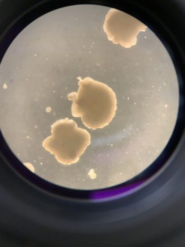
Through cutting-edge advancements in the field of genetics, researchers are now able to grow three-dimensional models of organs, called organoids, using in-vitro methods. Generating organoids for the human brain has become a pioneering research effort, especially the use of human embryonic stem cells (hESCs) to create human cortical organoids (hCOs). However, due to the fragility of these delicate models and the specific conditions required for growth, organoids can be quite difficult to generate and maintain for long periods of time. Hence, scientists continue to search for ways to optimize the generation of these 3D in-vitro models. One important aspect of organoid growth is the ability of blood to circulate both nutrients and oxygen efficiently. Recently published in Nature Methods, In-Hyun Park and his team at the Yale School of Medicine have discovered a way to induce vascular-like structures in hCOs that increase their functional maturation.
The key to inducing these vascular-like networks was the expression of the human E26 transformation specific variant 2 gene (ETV2). ETV2 over-expression enables the conversion of hESCs into endothelial cells, which line blood vessels. hCOs with 20 percent ETV2-infected cells were controlled to express ETV2 at a specific time point. “The remaining 80 percent were parental cell lines,” explained Bilal Cakir, first author of the paper. The different cells formed a 3D embryoid body after they were plated. “During the early stage of brain organoid development, the ETV2 expressing cells generated vascular-like structures within hCOs, called vhCOs,” Cakir said.
The researchers of this study performed several tests to analyze the process and effects of vascularization in vhCOs. “The most striking findings were that there is significantly less cell death after long-term culture in the vhCOs, and also that neurons become much more mature compared to control hCOs,” Cakir said. To study the effects of vascularization in vhCOs on organoid growth and cell death, he analyzed differences in size and growth rate of these organoids. He discovered that at day eighteen, before vhCO cells underwent ETV2 induction, the sizes of the organoids were similar. At day thirty and post-ETV2 induction, however, control hCOs were larger than vhCOs. But the growth rate of vhCOs increased over time, and at days 70 and 120, the two types of organoids had similar sizes again. Cakir believes that vascularization in vhCOs occurred around the time of their early growth lag and subsequent growth rate increase. In addition, vhCOs had significantly smaller amounts of cell death than control COs as the organoids continued to grow, suggesting that vhCO vascularization decreases cell death and enables organoid growth by carrying out oxygen diffusion.
To study how neuronal maturation and activity are influenced by ETV2 induction in vhCOs, Cakir performed whole-cell patch-clamp recordings to elucidate the action potentials produced by vhCOs and control hCOs. Results revealed that only one out of twenty control hCO cells produced any action potentials, while multiple action potentials were recorded for eight out of twenty vhCO cells. This data suggests vhCO vascularization enhances neuronal activity and maturation. Researchers also conducted gene set enrichment analyses that measured the resemblance between vhCO-derived neurons and in vivo developing human brain neurons. Results showed that vhCO-derived neurons were similar to gestational week 16-19 human neurons, whereas the corresponding control hCO-derived neurons were more similar to gestational week 10-12 human neurons. These results show that ETV2-induced vascularization increased the speed at which neurons were able to functionally mature in brain organoids.
Cakir’s team concluded that a functional vascular-like structure can be created in vhCOs through induced ETV2 expression that subsequently reprograms EC cells. Some key characteristics of this vasculature are its ability to decrease cell death and expedite functional neuron maturation. Despite these findings, Cakir believes that further optimization of brain organoid development is needed. “The vascularized organoids still need to be optimized to be used for even later stages,” Cakir said. In addition, he acknowledges the importance of brain organoids and their potential for studying various neural disorders: “Brain organoids hold the platform for investigating early stages of neuron wiring [and] are very critical for studying neuropsychiatric and neurodevelopmental disorders,” he said. ETV2-induced vascularization of brain organoids provides a platform for researchers to study these diseases at early stages, opening up a wide range of possibilities for future neurodevelopmental research.