
Image courtesy of www.pickpik.com.
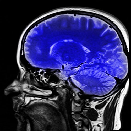
Attempting to peer inside the brain to diagnose neurological diseases such as Alzheimer’s Disease (AD) is complicated, requiring expensive equipment and expert analysis. Early detection of Alzheimer’s provides a range of benefits, including better treatment options for afflicted patients. While the standard technique for diagnosing Alzheimer’s has long been the analysis of Magnetic Resonance Imaging (MRI) scans, former Rochester Institute of Technology PhD student Viraj Adduru, working in conjunction with Geisinger Health Systems, has taken a step toward early diagnosis of this devastating disease by pioneering a cheaper and more accessible technique known as CTSeg.
The two primary brain imaging technologies are MRI and Computed Tomography (CT). MRI scans are more expensive and harder to acquire than CT scans, which presents a natural motivation for developing a technique involving CT scans to diagnose Alzheimer’s. The analysis of both MRI and CT brain images aims to calculate Total Brain Volume (TBV), which is an important indicator of AD. As Alzheimer’s progresses, brain matter deteriorates and TBV decreases. Image segmentation, where a human or program attempts to differentiate the brain tissue from the rest of the scan, is one approach to calculating TBV. The most common approach is having a trained specialist do this by hand, a process that can take 2-3 hours for a single brain image. Adduru’s automated method, CTSeg, is far more efficient. CTSeg uses a standard statistical template to compute a statistical map of the brain, where the probability stored in each voxel (like a pixel, but for 3D images) is the probability of that voxel being brain tissue. The final step of the algorithm involves converting that statistical map into a binary representation of the brain composition—tissue or not tissue. To generate the statistical map, Adduru adapted SPM (Statistical Parametric Mapping), which is a tried and true method used for the segmentation of MRI images, to work on CT images as well.
This project was inspired by the disparity between the number and quality of methods available for MRI analysis versus CT analysis. “In the MRI space there are so many methods…but in a hospital setting CT is more widely used, and there was no reliable method to estimate brain volume from CT images,” Adduru said. As he sought to address the problem, Adduru found that his experience in the hospital was a major reason why he was able to solve the problem so effectively. “The closer to the problem you are, the more inspired you are and you also come up with the methods that are most relevant and focused on solving the problem,” Adduru said.
The paper mentions the specific application of detecting group differences in TBV in a much broader sense. He hopes CTSeg can be used as a baseline for validating new models attempting to estimate TBV. With the increasing importance of machine learning, it is likely that Adduru’s CTSeg method will have an important place in medical research for years to come.