To study an animal’s brain in real-time as it navigates its surroundings, researchers typically implant electrodes into the animal’s brain. These electrodes track electrical changes in neurons, detecting when the neurons are activated. However, this method is limited in how many neurons can be analyzed simultaneously. “When you are looking at activity with electrodes, you are unsure if it is coming from one neuron or another area,” said Catherine Dulac, professor of molecular and cellular biology and researcher at Harvard University.
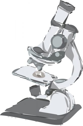
To procure a broader view of the brain, Dulac and her team have begun to used microscopes mounted on top of the heads of mice. “Typically, a microscope is something on your desk and you put a slide under the lens to look at tissue or fats,” Dulac said. “[But] this microscope is a miniature version–light enough to sit on the head of a mouse.” Small and portable, these miniaturized versions of fluorescent microscopes can sit on the head of mice, collecting live data while the mice perform various activities. These microscopes are connected to the neurons in the brain through an endoscopic tube, or a camera that peers inside an organism. Instead of tracking the change in electrical charge between neurons, researchers can now view the fluorescence of calcium binding in neurons, for example, to show signaling between neurons in the brain. To pass on a message, one neuron releases calcium into the synapse to activate signal receptors on the next neuron. The movement of calcium produces a change in fluorescent intensity that can be observed by researchers. This method allows researchers to visualize the activity of hundreds of thousands of neurons all at once, rather than focusing on just a few.
Dulac’s team has begun to use these portable microscopes to investigate the brain processes underlying the perception of social cues. The portability of these microscopes enables the research team to track the activity of the medial amygdala–the brain region that responds to social stimuli–of a mouse as it interacts with other mice. The scientists can then see how a mouse’s brain interprets the cues which determine its response. For example, the researchers compared the brain activity of a male mouse attempting to mate with a female mouse with an onlooking male mouse who decided to combat the mating mouse. These microscopes can also collect data to make predictions for other neurons. “You can take half of the neurons that you have recorded and model the form of activity mathematically to predict the activity in the other half,” Dulac said. “To some extent, you can simply look at neural activity and make a guess of what the animal is thinking and doing.”
In the future, researchers hope to apply these microscopes to image multiple areas at once. This function would give researchers the ability to see the dialogue between multiple areas of the brain, investigating how brain signals travel and pass between different brain regions.
Additionally, researchers are looking into a number of improvements, such as sharpening the spatial resolution of the microscope’s images to detect more fluorescent colors. Dulac’s team is also working on a smaller, potentially wireless version of the device as well as a multifunctional head-mounted microscope with an electrical probe to stimulate or inhibit neural activity. “For a neuroscientist, it’s a dream come true to have a window to what the brain does when an animal is looking around and making decisions,” Dulac added.

