
Imagine yourself mowing a lawn. If it was a leisurely Sunday morning, you might go back and forth in neat rows to ensure that every blade of grass was evenly cut. But what if you were in a rush? You might try more efficient paths such as spirals or sun spokes that leave small, uncut patches but quicken mowing time. Another option is to use a larger mower. This change yields better results regardless of the path chosen. With this rationale, a research team at the Magnetic Resonance Research Center at Yale developed a novel approach called Cartesian-Fast ROtary Nonlinear Spatial ACquisition (FRONSAC) and demonstrated its clinical potential. This technique improves magnetic resonance imaging (MRI) scan time and quality. The project was led by Sequence Developments Engineer and PhD candidate Nadine Dispenza and supervised by associate professor Gigi Galiana.
Getting up to speed: history of MRI
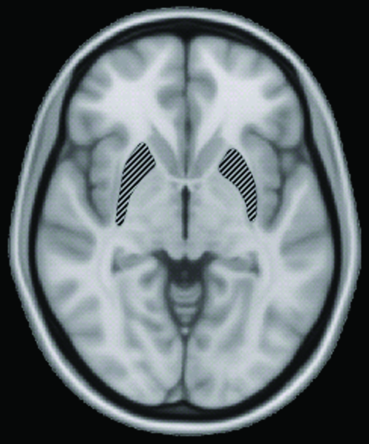
Used in medical diagnoses, MRI is a ubiquitous scanning technique that applies a magnetic field and sends radio wave pulses into the object of interest. The magnetic resonance signals, collected by receiver coils, are translated into a summation of sine waves via a mathematical process called Fourier transformation. In this way, the scanned object is not represented in real image space in the way we see the world, but rather in k-space, a representation of spatial frequencies in the image. With enough spatial frequencies mapped out, the k-space data can be transformed back into a real image that patients receive from their doctors.
Since the advent of MRI, linear magnetic field gradients—not unlike the conventional method of lawn mowing—have been used to sample k-space by acquiring one point at a time in a linear motion. This scanning method is called Cartesian imaging and is used for most clinical MRI. Though it is accurate, its long scan time makes the process expensive and limits the amount of data that can be collected. This limitation poses a problem for clinical imaging. “A lot of things don’t just rely on one black and white picture. You need a whole scan series,” Galiana said. From a clinical perspective, it is also undesirable to require patients to stay in the machine for long periods—it can be mentally and physically taxing for them, and their movements may interfere with data collection.
To accelerate scanning, parallel MRI was developed. Instead of sampling one k-space point at a time, coil arrays sample multiple differentially weighted points. That is, they measure a sampling distribution in k-space that is static in shape. In mowing terms, a wide mower is used that cuts grass shorter at its center than at its edges. Sampling in this way allows for gaps to be present in the data, as they will be filled by contributions from neighboring samples. This is analogous to using an array of lawnmowers and mowing in rows that are further apart, since the overlap at the edges of the lawnmower will eventually make the lawn even. This “undersampled” Cartesian imaging method shortens scan times. Unfortunately, it produces low quality images.
To improve scan time and image quality, research teams are exploring nonlinear gradient (NLG) encoding, which creates unique sampling distributions that dynamically change shapes within each line readout. Past studies have tested wide sampling distributions but did not yield considerable practical improvements. A better method would require a novel approach, and the Yale researchers were able to come up with a key piece of insight. “It’s not about how much of k-space we cover at once, but about how well we can resolve each unique point in k-space,” Dispenza said. The group had previously developed FRONSAC, a method that adds small perturbations of NLGs. By testing this method with Cartesian imaging, they hoped to skimp on the scanning and still obtain a high-quality image. The hope was that preserving overall linearity and adding small NLG perturbations to fill in the gaps could make MRI both fast and accurate.
Not so fast: research obstacles
Dramatic improvements in scan time appeared in the second iteration of the experiment. “It worked unusually quickly for research,” Dispenza said. By combining FRONSAC with tried-and-true standard Cartesian imaging, common problems associated with non-Cartesian imaging methods were avoided. The group showed that high-quality images can indeed be formed with relatively few samplings, thereby accelerating MRI.
The ride was not entirely smooth, however. The group had to first obtain a gradient coil, meaning they had to work with new hardware. Then, a few months after the first successful experiments, visual artifacts kept cropping up in k-space. The group spent months hunting down various possible causes, fearing that their success was not reproducible. They then noticed that artifacts appeared even with no sample in the magnet. Out of sheer desperation, the team modified the room humidity. In doing so, they noticed odd behavior with the equipment and shared their findings with colleagues at Siemens. Ultimately, it took half a year to identify the culprit: a broken capacitor. Hardware problems such as this are a common struggle in research, especially when pushing the boundaries of available equipment. “You’re always trying to do new things with new equipment, so these are the bumps that you have. It was helpful that when we spoke to people, we did not gloss over our hardware problems,” Galiana said. With the capacitor fixed, the rest was smooth sailing.
Cut to the chase: future directions
Since Cartesian-FRONSAC only requires small nonlinear perturbations in the magnetic field, this strategy can be implemented in preexisting hardware to shorten MRI scan times—all at a reasonable cost. Coils that generate these NLG fields may soon be in MRI scanners. Outside of clinical applications, this finding may also change industry standards. With Cartesian imaging, even slightly imperfect linear gradients would distort MRI images. Gradient linearity was therefore required to be perfect. The success of Cartesian-FRONSAC may allow for relaxation of industry standards regarding gradient linearity.
The research team is now further optimizing Cartesian-FRONSAC. For one, a protocol is in development to quantitatively determine exactly how much Cartesian-FRONSAC accelerates MRI. Another direction is to optimize the exact shape of the dynamic sampling distribution. To make Cartesian-FRONSAC universally accessible, Dispenza and Galiana are collaborating with another team to determine if NLG perturbations can be generated with radio frequency coils, which already exist in current MRI scanners. Finally, Cartesian-FRONSAC has thus far only been tested in two-dimensional imaging; experiments will soon push the envelope into three-dimensional imaging.
From a theoretical perspective, the team also hopes to further explore NLG encoding in k-space. This concept is foreign to many MRI researchers. “You can see the light-bulb moment [during conferences] where they think, ‘Wow, what other things could we have considered in k-space?’” Dispenza said. As future studies improve non-Cartesian imaging methods, FRONSAC can also be applied to accelerate these methods, especially as FRONSAC has already been proven to enhance the time efficiency of one such method, called turbo spin echo (TSE) imaging.
The key has been put into the ignition to unlock the potential that FRONSAC holds for improving clinical imaging. It’s time to start mowing lawns.