
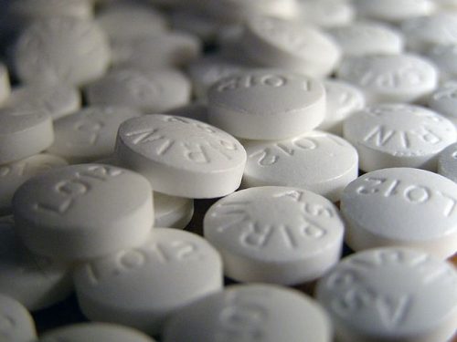
In communities across America, the opioid crisis is ripping apart families. Overdoses are leaving children orphans and addiction is ruining lives. The root cause of the issue is the over prescription of opioid painkillers by medical professionals. Between 21% and 29% of people who are prescribed opioids for chronic pain misuse them, and 80% of heroin users started by misusing opioids. The opioid epidemic has become a widespread public health crisis in the United States. Beyond increases in opioid misuse and overdoses, the increase in injection drug use has caused a resurgence of diseases like HIV and hepatitis C. Stephen Waxman’s lab at Yale University is looking to change this by developing safe and non-addictive treatments for chronic pain.
Scientists in Waxman’s lab, led by postdoctoral associate Liz Akin and senior research scientist Suleiman Dib-Hajj, are taking steps towards this goal by visualizing the specific pathway of pain for the first time. Their focus was on Nav7.1 sodium ion channels, which Dr. Akin describes as “volume knobs for pain.” Researchers have demonstrated that enhanced expression of Nav7.1 occurs after injury and that more channel activity causes more pain. This channel is found in the axonal endings of nociceptors, a type of sensory neuron located throughout the body.
Dr. Akin and Dr. Dib-Hajj discovered that Nav1.7 channels are transported along microtubules in vesicles to the cellular membrane in the distal axon surface. This process is an example of conventional vesicle trafficking. Previously, scientists have tried to study this pathway by tagging the channels with green fluorescent protein (GFP), however, only a few moving vesicles were able to be observed with this strategy, so it was hard to draw conclusions. Dr. Akin and Dr. Dib-Hajj developed a new imaging technique, called OPAL imaging, which is a pulse chase method of fluorescently labelling Nav1.7 vesicles. Through this method, they showed that channel transport was microtubule dependent. They treated the cells with miconazole, a microtubule destabilizing agent, and used OPAL imaging to observe how the movement of the vesicles was effected. They were able to determine the destination of the vesicles by showing that an hour after labeling, the Nav1.7 vesicles were concentrated at the distal end of the axons. They again used OPAL imaging to demonstrate that this pathway is an example of conventional vesicular trafficking by identifying the Rab protein associated with Nav1.7. Rab proteins are markers for vesicular trafficking. They cotrafficked Nav1.7 with eight different types of Rab and showed that the channel and Rab6A moved together, suggesting that they are packaged in the same vesicle.
Multiple drug companies have already been attempting to create therapies that target the current produced by the NaV1.7 channels. Many have been developed and have undergone clinical testing, but none have made it to the market. There is already a precedent for targeting ion channels in drug therapies. Lyrica is a current drug that targets calcium channels to treat epilepsy and nerve pain. When asked about the pharmaceutical application of this research Dr. Dib Hajj said “what we are proposing, now that we have a better understanding of the trafficking of this channel and how it gets deployed to the nerve endings where it exerts its function, is that it itself could become a target for drug development.” This could happen in a number of ways. A drug could affect NaV1.7 channels directly, which is how Lyrica affects calcium channels. Alternatively, once the lab identifies key genetic regulators of the trafficking pathway, they could use genetic therapies to control the process.
Dr. Akin and Dr. Dib Hajj’s work is especially instrumental because it develops a whole new way of looking at these processes in the form of OPAL imaging. In the words of Dr. Dib Hajj, “This lays the groundwork for looking at these types of problems.” Moving forward, Waxman’s lab is using this imaging technique to visualize the trafficking of other identified human pain channels and to focus on some of the genetic targets that they have already identified. They are also beginning to study the relationships between different pain channels to determine if their activity is individual or interconnected. Additionally, OPAL can be used to visualize any membrane proteins, so it can be used by scientists in other labs working on a variety of topics. This research expands the possibilities for pain research, bringing science one step closer to an effective, non-addictive alternative to opioids. This will be key in turning the tide of the opioid epidemic.