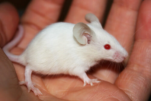
Image courtesy of Flickr.
Historically, in vivo mice models have fallen short of simulating human erythropoiesis, or the production of mature red blood cells (RBCs), because of mice’s immune response. As a result, human blood disorders such as sickle cell anemia have been mostly studied with murine RBCs from mice, which does not allow as much flexibility in experimentation. Recently, however, researchers from the Yale Department of Immunology and Yale Cancer Center have developed a murine model that displays enhanced human erythropoiesis and mature RBC survival.
This model relies on cytokine and liver humanization—the adoption of human genes and tissues—in order to extend the lifespan of circulating human RBCs in the mice. Without humanization, engrafted human RBCs can be detected by the mices’ immune systems and subsequently destroyed by immune cells.
Through the deletion of the fumarylacetoacetate hydrolase (Fah) gene and transplenic injection of adult human hepatocytes, the researchers succeeded in liver humanization. Fah gene deletion allowed the engrafted human hepatocytes to repopulate in an immunodeficient liver. Essentially, the researchers grew a human liver in mice.
Liver humanization reduced the production of a protein called mucin 3A by mice hepatocytes. Mucin 3A, when attached to human RBCs, mediates the adherence to mice phagocytes, which results in the destruction of the human RBCs. “Combining a humanized liver with a humanized blood system increased RBC count drastically,” said Richard Flavell, Yale professor and one of the study’s corresponding authors.
Yale researchers also succeeded in cytokine humanization by utilizing MISTRG mice, which carry human genes that encode cytokines such as interleukin-3, granulocyte-macrophage and macrophage colony-stimulating factor, and thrombopoietin. These cytokines, in addition to a protein called signal-regulatory protein alpha, regulate innate immune cell development and human blood cell development in mice. Liver and cytokine humanization therefore enhances the survival of engrafted human RBCs by preventing the mice’s immune responses from targeting the foreign human cells.
This mouse model saw success in simulating sickle cell disease as well. Sickle cell anemia in humans causes red blood cells to contort into a sickle shape, which can block blood flow and restrict oxygen and nutrient supply to the body. The researchers observed that after engraftment of hematopoietic stem and progenitor cells from sickle cell patients, the mice started producing sickle-shaped human RBCs. Furthermore, they started displaying signs of sickle cell anemia in their lungs, liver, kidneys and spleen, such as alveolar hemorrhage, thrombosis, and vascular occlusion. The sickle cell studies in mice showed that this model can be adapted to studying other RBC disorders, including thalassemia, and even hematopoietic stem cell diseases like myelodysplasia and erythroleukemia.
“We also like to think of this model as one that potentially addresses health disparities. We hope that it will allow us to advance treatments for sickle cell disease, a disease predominantly affecting African Americans in the US,” said Yale professor and corresponding author Stephanie Halene. “Now that we’ve succeeded in growing mice with human blood, future research may make huge breakthroughs in studying blood disorders that disproportionately affect groups of people.”
Citations:
Song, Yuanbin, et al. “Combined Liver–Cytokine Humanization Comes to the Rescue of Circulating Human Red Blood Cells.” Science, vol. 371, no. 6533, 2021, pp. 1019–1025., doi:10.1126/science.abe2485.

