
Image Courtesy of Pixabay.
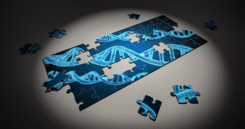
Let’s turn the clock way back to when you consisted of only a small clump of cells. How does this tiny clump of cells know to transform into various cell types throughout your body, from hair and eyes to lips and legs? It turns out that these instructions are encoded in our genome. However, our genome is initially “silent.” It needs to be activated and reprogrammed through a process called zygotic genome activation so that our cells can properly differentiate into specific cell types in our body.
The Giraldez lab at Yale specifically investigates the cellular mechanisms of this process, which involve a variety of interactions between protein ‘factors’ and DNA that work together to transform the “silent” state of the genome into the activated state. “It’s kind of like erasing the blackboard and writing new instructions,” said Antonio Giraldez, the Fergus F. Wallace Professor of Genetics. “We’re trying to understand the factors that erase the previous instructions and the factors that instruct the genome to employ the first cascade of events that will lead to development.”
Traditionally, researchers have relied on indirect biochemical methods to unravel the intricacies of this process, as the existing visualization techniques have presented considerable limitations. “We wondered if we could actually come up with a new way to visualize what happens inside of these clusters,” said Mark Pownall, a member of the Giraldez lab who first looked into this research direction. In collaboration with the Bewersdorf lab at Yale, Pownall adapted their already-established technique of pan-expansion microscopy (pan-ExM), incorporating labeling of protein, RNA, and DNA to create Chromatin Expansion Microscopy (ChromExM). Pan-expansion microscopy involves fixing cells to an expandable gel called a hydrogel that swells via addition of certain functional groups. By layering multiple hydrogels on top of each other, the cells take on a larger size that can be better resolved under a microscope.
The cell, and specifically the chromatin that makes up our chromosomes, expands to become four thousand times larger, allowing for a never-before-seen look into the small-scale interactions that thus far, researchers have only been able to theorize about. “This has allowed us to start measuring distances that are ten times higher in resolution than what we have ever captured before,” Giraldez said. “This has revealed how the models and the cartoons we are drawing from biochemical experiments can be visualized for the first time.”
One of the main concerns of expansion microscopy was whether the cell’s proportions could be maintained during the expansion process. To ensure that the cell expanded proportionally, the Giraldez lab developed a technique where the initial cell was marked with parallel stripes, and if the stripes remained parallel after expansion, it would have exhibited proportional growth. “This showed us that we preserved these stripes really well after they were cleaved, which was one hint that we actually preserved chromatin structure,” Pownall said.
With this novel technique in hand, the Giraldez lab could finally visualize the specific factors involved in activating the genome. Using ChromExM, they were able to study the function of Nanog, a protein that binds to a “gene enhancer” region of DNA that stimulates the activation of genes involved in development. Although Nanog had been shown to interact with RNA polymerase, located at the promoter region of the gene where the polymerase would initially bind to initiate transcription of DNA to mRNA, it was unclear whether these structures actually required transcription to form. By using ChromExM, they found that the Nanog protein was initially in close contact with RNA polymerase, but once transcription was initiated, the protein separated itself from the growing strand of mRNA. The Giraldez lab termed this interaction the “kiss-and-kick model,” where transcription acts as the “kick” to separate the enhancer and promoter regions.
The development of the human body is an incredibly complex process directed by genetic codes regulated by countless factors that interact with chromatin and different organelles in our cells. The novel technique of ChromExM not only allows us to visualize these processes, but is also accessible and applicable to other fields of study, as it only requires a confocal microscope that can be found in many research labs. “I think that this could add a fundamental tool to the global toolbox to really understand how different molecules interact in the nucleus by visualizing these fundamental processes of life,” Giraldez said. “The accessibility is great, and the possibilities to apply ChromExM to other approaches is very large.”