
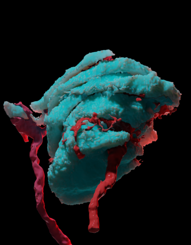
Image courtesy of Yury Nikolaev.
A light brush across the skin, the vibration of your phone in your hand when you receive a text, the sharp jab when you accidentally poke yourself with a pencil—we constantly receive various tactile stimuli, but how do our bodies sense these different types of touch? The skin contains many kinds of mechanoreceptors, which are cells or minuscule organs that respond to different intensities of pressure and frequencies of vibration. However, the detailed mechanism of touch detection by mechanoreceptors has not yet been elucidated.
To bridge this gap, Yury Nikolaev, Luke Ziolkowski, and colleagues in the Bagriantsev and Gracheva labs at Yale University used microscopy to resolve the structure of a type of mechanoreceptor called Meissner’s corpuscles in mallard ducks. Their findings point towards a novel bicellular mechanism of touch detection, in contrast to the canonical, single-cell-type-mediated mechanism.
Vertebrates detect touch in the skin using tiny organs called mechanosensory corpuscles, which contain sensory neurons and glial cells. Sensory neurons detect stimuli by sending electrical signals called action potentials, while glial cells provide structural support for neurons. Meissner’s corpuscles are a type of mechanosensory corpuscle responsible for detecting low-frequency vibrations and fine touch, and they include both sensory neurons and a type of glial cell called lamellar cells. Stimulating the sensory neurons within Meissner’s corpuscles transmits a signal to the brain that is interpreted as the sensation of touch.
The researchers used a cutting-edge microscopy technique called enhanced focused ion-beam scanning electron microscopy (FIB-SEM) alongside machine learning to resolve the structure of mallard duck Meissner’s corpuscles, and they found an unexpected structure. “We are the first to reveal the structure of any corpuscle using this technique,” Nikolaev said. The corpuscle contains stacks of lamellar cells integrated among sensory neurons. Typically, only sensory neurons are thought to be capable of detecting touch, but previous work done by Nikolaev indicated that these lamellar cells were responsive to mechanical stimuli. This prompted the researchers to investigate whether lamellar cells could trigger action potentials in the sensory neurons, thereby acting as secondary touch sensors. “There was some prior evidence from other types of nerve endings where there were occasionally non-neuronal cells that were detecting touch and helping communicate that to the sensory neuron,” Ziolkowki said.
When researchers triggered action potentials in lamellar cells, they found that the sensory neurons also transmitted action potentials. In contrast to direct stimulation of sensory neurons, which produces consistently timed action potentials, lamellar cell-induced neuron firing time varies considerably. This variability indicates a unique role for lamellar cells in touch sensing, possibly allowing for more sensitive and specific touch sensing.
While this research was performed on ducks, their Meissner corpuscles are very similar to those of other mammals, so it is likely that humans have a similar bicellular system involving cooperating lamellar cells and sensory neurons. Nikolaev and Ziolkowski will continue investigating the nature of the interactions between lamellar cells and neurons, challenging the paradigm of a simpler, unicellular model for touch detection and expanding our understanding of how we use touch to perceive the world.