
Art by Malia Kuo.
Scientists learn more about the cell every day. From taking microscopic pictures to performing biochemical tests on pellets of harvested cells, biologists are able to determine the organization and interactions of major cellular components, including organelles, proteins, and nucleic acids. But understanding what these interactions look like in real-time is much more difficult than capturing fluorescent images, which offer a freeze-frame snapshot of the cell, and biochemical assays, which offer only a general understanding of molecular interactions but are often limited by the experimenter’s ability to manipulate the reacting biomolecules in space and time. In a recent article in Science Advances, Yale Professor of Cell Biology and Biomedical Engineering Chenxiang Lin and postdoctoral fellow Longfei Liu pioneer a unique way to study cell biology by harnessing the power of DNA as a molecule with a highly controllable structure to study the interactions of proteins and the cell membrane in real-time.
Students typically learn that DNA is the genetic code of the cell, responsible for encoding the information eventually converted to the proteins responsible for cellular functions. But Lin offers an alternate perspective on DNA: that DNA itself has unique structural and chemical properties that can guide the assembly of other biomolecules and modulate how they interact. The DNA contained within our cells’ nuclei is in the traditional double-stranded helix because this conformation keeps DNA stable and relatively easy to transcribe. But scientists can now control the sequence of short single-stranded DNA molecules, called oligonucleotides, such that they spontaneously form nanoscale assemblages of precisely defined shapes. And because DNA oligonucleotides are easier to synthesize and chemically modify than molecules not found in nature or even proteins, they form nanostructures desirable for biochemical and biophysical experiments, where scientists want to study the finest details of molecular organization, dynamics, and function. The programmability and self-assembling nature of the DNA structures allow scientists to repeat such experiments many times and with all kinds of permutations. “This bottom-up approach is very powerful since all you need to do is design the DNA molecules correctly. The DNA strands can find each other and self-assemble into larger structures, with precise experimenter control.” Lin explained.
History of DNA Nanotechnology
According to Lin, the idea to use DNA as a structural macromolecule for more than just encoding genetic material harkens back forty years to New York University Professor Ned Seeman. Seeman imagined DNA nanostructures completely conceptually before creating them was even possible. DNA nanotechnology began to materialize upon creating a stable four-way DNA junction in a test tube resembling the Holliday junction, a somewhat complicated three-dimensional arrangement of single-stranded DNA pieces that forms during a type of DNA repair called homologous recombination. Scientists then began experimenting with combining multiple small, single-stranded DNA oligonucleotides into “tiles” and harnessing the symmetry of these tiles to build two-dimensional lattices or three-dimensional crystalline structures. In 2006, Paul Rothemund at the California Institute of Technology invented a new technology called DNA origami. He folded a single-stranded DNA extracted from viruses into shapes like smiley faces with the help of tens to hundreds of oligonucleotides. Importantly, these helper strands could each carry additional modifications to attach other molecules to the DNA origami structure at precise locations.
Scientists sought to create domains in the DNA nanostructure that would change in conformation in response to some physiologically relevant signal, such as changes in pH, visible light, or UV radiation. These DNA nanostructures can mimic proteins to study how changes in these proteins’ structures would impact their biochemical activities. DNA structures containing regions of non-conventional motifs, such as four stacked cytidine bases (one of four nitrogen-containing cyclic molecules that comprise the inside of the DNA helix), change in conformation in acidic environments, which are characteristic of cancerous cells. Similarly, chemically joining an azo-benzene group (two hexagonal carbon rings connected by two nitrogen atoms) to the DNA backbone allows DNA to change conformation in response to a visible or UV light source. Such trigger-responsive DNA structures allow engineers to build nanorobots under users’ command by adding a drop of a chemical or simply by shining a light. This is also very useful for mimicking the dynamic activity of some proteins that act as enzymatic molecular switches.
Studying Membrane Dynamics
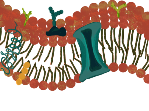
Unfortunately, while DNA nanotechnology has seen some traction in studying soluble, cytoplasmic proteins, proteins embedded in the cell’s membranes are harder to manipulate. Membrane proteins are crucial for many of the cell’s most basic functions, such as motion, regulating molecular traffic through the membrane, and interacting with pathogens, all of which require the membrane proteins to respond to changes in the membrane landscape and sometimes actively remodel the membrane. However, the membrane proteins are often involved in very complex interactions with lipids, other membrane proteins, and the cytoskeleton, scaffolding proteins that give cells their shape. Because this system is so complicated, Lin and Liu aimed to build a highly reductionist cell membrane model to contain only some features or proteins of interest. In their article, the proteins were foregone and replaced with mimics made of DNA. “These DNA structures were designed to look like a membrane-remodeling protein and work like one,” Lin said. “By tweaking them and observing how they behave on membrane, we may learn a thing or two about how the protein works in cells.” This system enabled them to study cell membranes as a platform and create artificial environments relevant to biological problems.
One of the most important membrane dynamics researchers have attempted to model is membrane tubulation, whereby one part of a membrane pokes out from an existing piece of a membrane but remains in close contact, essentially creating an extended “tube” of the membrane. This process is ubiquitous across the cell, involved in organelle and cell division, as well as packaging molecules for transport in membrane-enclosed vesicles. The interaction requires exquisite molecular machinery to change the membrane curvature and actually pinch the membrane destined to be separated from the old membrane (budding).
Liu and Lin were able to understand the key factors required for membrane vesicle formation to occur using DNA nanostructures. The DNA nanostructures in their study mimicked the proteins that integrate into the membrane, contributing to membrane curvature. This allowed them to study the proteins’ properties and their effect on tubulation and vesiculation. The DNA nanostructures they integrated into their cell membrane model were initially set in an open state with high internal tension. Releasing such tension caused the DNA structures to buckle and adopt a closed, highly curved conformation (imagine a spring-loaded clamp). When they operated such DNA nano-clamps on the membrane, membranes were able to be curved, and many DNA-coated membrane tubes spontaneously emerged. However, fewer tubes were observed when the closed DNA clamps were less curved themselves. Moreover, releasing the DNA clamps from the high internal tension state seemed important for tubulation since preventing the DNA from changing from its initial state prevented the phenomenon. Interestingly, removing the DNA from membrane tubes led to vesiculation. Thus, Liu and Lin were able to deduce that the curvature of certain membrane proteins contributes to membrane curvature and that the ability of these proteins to switch conformation can release energy and act as a biophysical switch to determine whether the membrane could bud or not.
Next Steps
Lin and Liu’s study was landmark in several ways. They were able to provide a proof of concept that the geometric properties of DNA nanostructures could act as a reliable substitute for the geometric properties of a membrane-bound protein and that tuning the mechanical properties of the DNA structures to modulate membrane dynamics could provide insights into how proteins impact membrane dynamics. “Membrane proteins are quite hard to manipulate. With DNA nanostructures, we can control the structure, shape, geometry, and modifications, which means we can do experiments in a more controlled way,” Liu said. Moreover, they were able to model membrane tube formation across multiple designs and experimental conditions—such as by varying the curvature and internal tension of the DNA clamps and changing the starting curvature of the membrane—and show that the curvature of the DNA nanostructure was the instigating factor in tubulation across these varying conditions. This highlights the unique advantages of using DNA nanostructures for such experiments. These nanostructures enable researchers to tune previously inaccessible parameters and ensure experiment reproducibility.
However, both Lin and Liu expressed some caution about applying such conclusions definitively to the cells of living organisms. First, while DNA nanostructures may be used to structurally approximate proteins, key differences in the two classes of molecules’ stability, folding, and activity need to be accounted for when making conclusions about how proteins interact with membranes. Secondly, since the cellular models they employed were simplified, their conclusions from their experiments will probably need to be verified by studies in cells extracted from living organisms since membrane proteins in cells are engaged in many more interactions than the minimal set. Both researchers are embarking on new projects, including several in collaboration with Martin Schwartz, a Yale professor, to study how membrane proteins and the DNA structures that mimic them act on membranes with underlying cytoskeleton in major cellular processes.
Lin and Liu aim to further investigate how external signals impact DNA nanostructures mimicking membrane proteins and how cellular processes are accordingly modulated. Eventually, they aim to harness the similarities between membrane proteins and DNA nanostructures to create a reliable cell model from scratch. “It would be very cool to build synthetic cells that would work similarly to naturally existing cells,” Lin said. With these headways in the field of DNA nanotechnology and cell biology, the scientific community is on its way to learning more about real-time cellular processes than ever before.